

X
Trustworthy Source
National Health Service (UK)
Public healthcare system of the UK
Go to source
Understanding Intravaginal Ultrasound
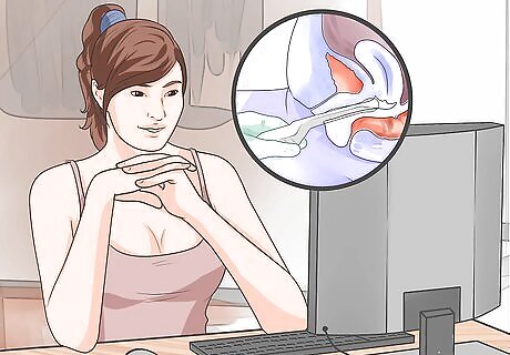
Understand what an intravaginal ultrasound is. Intravaginal ultrasound is used for visualizing the organs inside your pelvic area. It can be used to diagnose gynecological conditions (such as pelvic pain and unusual bleeding) or visualize the early phases of pregnancy. During the procedure, your doctor inserts a transducer, which is about the size of a speculum, into your vagina. From there, the transducer emits sound waves that allow your doctor to visualize your internal organs. Intravaginal ultrasounds are painless but you might feel pressure and discomfort during the procedure.

Know if you need an intravaginal ultrasound. Intravaginal ultrasounds are performed whenever your doctor needs to take a closer look at your reproductive organs, such as the cervix, ovaries, and uterus. Your doctor may also perform an intravaginal ultrasound to monitor your pregnancy and fetus. Your doctor may order the procedure if you experience unexplained pain, bleeding or bloating. For example, intravaginal ultrasounds can reveal changes in the shape and density of your reproductive tissues and can also be used to visualize blood flow to your pelvic organs. It can be used to monitor fibroids, ovarian cysts, and cancerous growths in your pelvic organs or diagnose the cause of vaginal bleeding and cramping. Intravaginal ultrasound can also help diagnose infertility issues or abnormalities in the bladder, kidneys and pelvic cavity. During pregnancy, your doctor can use it to detect the early stages of pregnancy, monitor the development of your fetus, detect multiples, and rule out an ectopic (tubal) pregnancy.
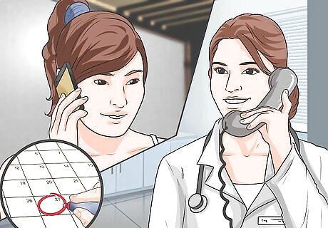
Schedule the procedure. The timing of this procedure depends on the reason why you need to have it done. During pregnancy, an intravaginal ultrasound can occur as early as 6 weeks after conception but typically is done between 8 and 12 weeks of pregnancy. If your doctor is trying to diagnose a reason for unusual pain or bleeding, your procedure will likely be scheduled immediately. If you need an intravaginal ultrasound for infertility issues, then your doctor may choose to perform it around the time you ovulate. An intravaginal ultrasound can be performed at any time during the menstrual cycle, but it is usually best to have it done right after your period ends, between days 5 and 12 of your cycle. During these days, your endometrial lining is the thinnest, which allows for clearer images of your uterus.
Preparing for the Ultrasound

Take care of personal hygiene before you leave your home. You will want to take a shower/bath prior to leaving your home to have your intravaginal ultrasound. If you are on your menstrual cycle and you are wearing a tampon, you must remove it before the procedure. Make sure you bring an extra tampon (or feminine napkin) with you to use after the procedure.

Wear comfortable clothing that is easy to remove. You will need to wear a hospital gown for the procedure so it is a good idea to wear comfortable, easy-to-remove clothes. You should also wear shoes that are not difficult to take off as you will need to remove clothing from waist down. Sometimes you can keep your clothing from waist up so you may want to wear separates instead of a dress.

Ask your doctor whether you should empty your bladder. Typically you should have an empty bladder for the procedure. Use the restroom just before the procedure and do not drink anything for 30 minutes prior to your intravaginal ultrasound. Sometimes your doctor may perform a transabdominal ultrasound first. For this, a partially full bladder is preferred as it lifts the intestines and allows your doctor to see the pelvic organs more clearly. If your doctor asks you to have a partially filled bladder, you will need to drink water prior to the ultrasound and not use the restroom. You should begin drinking water a half an hour before your ultrasound. You might be asked to empty your bladder before the intravaginal ultrasound.

Fill in any necessary paperwork. Once you get to the hospital or clinic, you must sign a consent form stating that you agree to have the intravaginal ultrasound performed. Also let your doctor know if you have a latex allergy. The transducer is covered with a latex or plastic sheath before it is inserted into the vagina.
Getting the ultrasound

Change into the gown provided. Once you are taken into a dressing room or the ultrasound room, remove your clothes and change into a hospital gown. Sometimes you need to undress from the waist down only. In this case, you will typically receive a sheet to use as a cover during the procedure.

Lie on the bed. After you have undressed, go and lie down on the examination table. Intravaginal ultrasounds are performed while you are lying on your back similar to when you receive a normal gynecological exam. You need to bend your knees and rest the soles of your feet flat on the stirrups connected to the examining room bed in order to give your doctor the best access to your vagina.
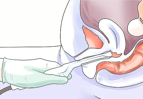
Allow your doctor to insert the transducer. Before inserting the transducer, your doctor will place a plastic or latex sheet over it and lubricate it with a gel to allow for easier insertion. Your doctor will then gently insert the transducer into your vagina in order to begin constructing the image. The transducer is slightly bigger than a tampon and is designed to fit into your vagina comfortably.
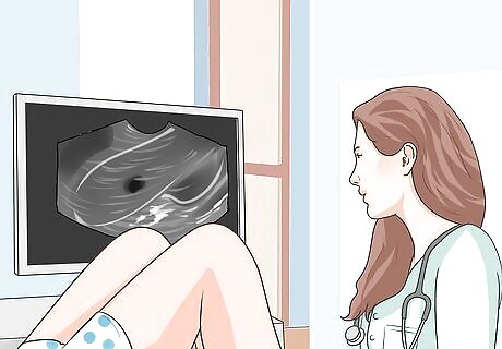
Know what to expect during the procedure. Your doctor holds the transducer inside of your vagina and may rotate it slightly in order to create a clear image of your pelvic organs. The transducer is connected to a computer. Once inserted, images of your pelvic organs will begin to appear on the computer screen. Your doctor will check the screen throughout the scan to make sure that everything is showing up in detail. Your doctor may also take pictures and/or live video. If the ultrasound is performed to monitor your fetus, your doctor typically prints the pictures and gives them to you.

Clean up and get dressed again. Intravaginal ultrasound typically last no longer than 15 minutes. After the procedure is over and your doctor removes the transducer, then you are given privacy to dress. You will be given towels to remove any gel that remains on your inner thighs and/or pelvic area. You may also want to visit the restroom in order to wipe excess lubricant from your vagina and insert a new tampon.

Ask about the results. If your primary doctor performs the ultrasound, she may describe preliminary results as they appear on the screen. If you are referred to a different clinic, then you usually must wait for your primary doctor to receive a written report of your results. You receive the results of your scan based on the complexity and urgency of your condition. If your ultrasound is strictly exploratory, then you will likely wait a few days to a week for your results.


Comments
0 comment